Arthroscopy of the Knee Joint
Knee Arthroscopy is a common surgical procedure. It is performed using an arthroscope, a viewing instrument, to look into the knee joint to diagnose or treat a knee problem. It is a relatively safe procedure and a majority of the patient’s discharge from the hospital on the same day of surgery.
Knee anatomy
The knee joint is one of the most complex joints of the body. The lower end of the thighbone (femur) meets the upper end of the shinbone (tibia) at the knee joint. A small bone called the patella (kneecap) rests on a groove on the front side of the femoral end. A bone of the lower leg (fibula) forms a joint with the shinbone.
To allow smooth and painless motion of the knee joint, the articular surfaces of these bones are covered with a shiny white slippery articular cartilage. Two C-shaped cartilaginous menisci are located in between the articular surfaces of the femur and tibia.
The menisci act as shock absorbers providing a cushioning effect to the joints. Menisci also play an important role in providing stability and in load bearing.
Bands of tissue, the capsule, and the cruciate and collateral ligaments, keep the different bones of the knee joint together and provide stability to the joint. Surrounding muscles are connected to the knee bones by tendons. This ligamentous capsule is lined with a synovial membrane that secretes synovial fluid for lubrication.
Indications for Knee arthroscopy
The knee joint is vulnerable to a variety of injuries. The most common knee problems where knee arthroscopy may be recommended for diagnosis and treatment are:
- Torn meniscus
- Torn or damaged cruciate ligament
- Torn pieces of articular cartilage
- Inflamed synovial tissue
- Misalignment of patella
- Baker’s cyst: a fluid filled cyst that develops at the back of the knee due to the accumulation of synovial fluid. It commonly occurs with knee conditions such as meniscal tear, knee arthritis and rheumatoid arthritis.
- Certain fractures of the knee bones
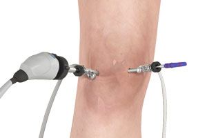
Procedure
Knee arthroscopy is performed under local, spinal, or general anaesthesia. Your anesthetist will decide the best method for you depending on your age and health.
- The surgeon makes two or three small incisions around the knee.
- Next, a sterile saline solution is injected into the knee to push apart the various internal structures. This provides a clear view and more room for the surgeon to work.
- An arthroscope, a narrow tube with a tiny video camera on the end, is inserted through one of the incisions to view the knee joint. The structures inside the knee are visible to the surgeon on a video monitor in the operating room.
- The surgeon first examines the structures inside the knee joint to assess the cause of the problem.
- Once a diagnosis is made, surgical instruments such as scissors, motorized shavers, or lasers are inserted through another small incision, and the repair is performed based on the diagnosis.
The repair procedure may include any of the following:
- Removal or repair of a torn meniscus
- Reconstruction or repair of a torn cruciate ligament
- Removal of small torn pieces of articular cartilage
- Removal of loose fragments of bones
- Removal of inflamed synovial tissue
- Making small holes or microfractures in the damaged articular cartilage to stimulate cartilage growth
- After the repair, the knee joint is carefully examined for bleeding or any other damage.
- The saline is then drained from the knee joint.
- Finally, the incisions are closed with sutures or steri-strips, and the knee is covered with a sterile dressing.
After the Surgery
Most patients are discharged the same day after knee arthroscopy. Recovery after the surgery depends on the type of repair procedure performed. Recovery from simple procedures is often fast. However, recovery from complicated procedures take a little longer.
Pain medicines are prescribed to manage pain. Crutches or a knee brace may be recommended for several weeks. A rehabilitation program may also be advised for a successful recovery. Therapeutic exercises aim to restore motion and strengthen the muscles of the leg and knee.
Risks and complications
Knee arthroscopy is a safe procedure and complications are very rare. Complications specific to knee arthroscopy include bleeding into the knee joint, infection, knee stiffness, blood clots or continuing knee problems.